Eye Health Screening Evaluations
We continue to invest in traditional examination equipment – items that use light and magnification to examine the eyes inside and out. However, we have also embraced technology that “scans” the eye and provides much MORE information for our doctors to use in evaluating your eye health. This technology is typically not covered by insurance. There is an additional charge of $17-$49 for these images.
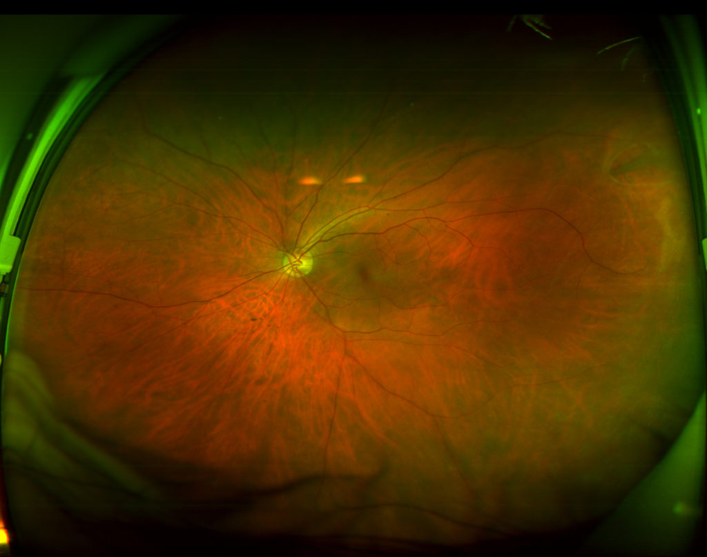
The Optomap retinal imaging unit has been part of our practice since the early 2000s. This “looking in” view has become an indispensable part of a complete exam.
In addition to providing extra capabilities to ascertain the “level” or “layer” of a problem, the real power in the images comes when we have annual – year after year – images to compare with each other.
The images below were from a patient with no problems in his vision. Immediately, a “horseshoe tear” to the retina was observed in the right, slightly upper area (about 2:00). The patient was scheduled with a retina specialist, and laser treatments were applied to prevent this from leading to a loss of vision (2nd image – after treatment). Optomap images are done on patients of all ages.

The iWELLness retinal exam is a cross sectional view of your retina. It reveals changes in retinal anatomy that may not be seen by traditional methods. It is most appropriate for those 18 years old and above.
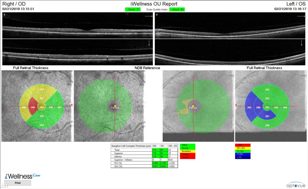
The first set of scans above demonstrate a “cross-section” of the central part of our retina – the macula – your doctor will review the individual layers for thickness and irregularity. This is a great way to diagnose early stages of macular degeneration.
The second set of scans helps us to identify changes related to both retinal and nerve fiber layer disease. These scans check for thickening or thinning of the retina which can indicate diseases such as glaucoma and degenerative nerve disorders such as Alzheimer’s. These changes can not be identified with traditional eye exams. Finding changes early allows us to begin management YEARS before than if we waited for other glaucoma damage.
We believe that BEST CARE of your eyes includes annual retinal imaging as outlined above.
If you have any questions about retinal imaging, please ask your doctor at your next visit.